Uvea

The uvea is the middle layer of the eye. It lies beneath the white part of the eye (the sclera). It is made of the iris, ciliary body, and choroid. Inflammation of one or more of these structures is called uveitis.

What is Uvea?

The uvea is the middle layer of the eye. It lies beneath the white part of the eye (the sclera). It is made of the iris, ciliary body, and choroid. These structures control many eye functions, including adjusting to different levels of light or distances of objects

Symptoms Of Uvea

Pain in the eye

Redness of the eye

Blurred vision

Photophobia

Floaters.

Pain in the eye

Redness of the eye

Blurred vision

Photophobia

Floaters

Cause Of Uvea

Infections (Bacterias, Viruses Fungus and protozoal)

Auto immune diseases like Ankylosing spondylitis, Rheumatoid arthritis, Psoriasis, SLE, Behcet’s syndrome, Sarcoidosis, VKH disease etc.,

Cancers can cause uveitis.

Trauma can cause uveitis in the same eye or even other eye

When the cause is unknown the uveitis is known as idiopathic.

Infections (Bacterias, Viruses Fungus and protozoal)

Auto immune diseases like Ankylosing spondylitis, Rheumatoid arthritis, Psoriasis, SLE, Behcet’s syndrome, Sarcoidosis, VKH disease etc.,

Cancers can cause uveitis.

Trauma can cause uveitis in the same eye or even other eye

When the cause is unknown the uveitis is known as idiopathic

Uveal tract

The prime functions of the uveal tract as a unit are

Are You Prone To Vision Loss
Due To Uveitis?

Complete Uvea Treatment With State-Of-The-Art Technology

The binocular slit-lamp examination provides a stereoscopic magnified view of the eye structures in detail, enabling anatomical diagnoses to be made for a variety of eye conditions. A second, hand-held lens is used to examine the retina. A slit-lamp exam is usually done during a regular checkup with your eye doctor before the cataract surgery procedure.

NC Tonometer is used to perform Tonometry. Tonometry is a quick and simple test that checks the pressure inside your eyes. The results can help your doctor see if you're at risk for glaucoma. The pressure inside your eye is called intraocular pressure (IOP).

This test measures fluid pressure in your eye. The test involves using a slit lamp equipped with forehead and chin supports and a tiny, flat-tipped cone that gently comes into contact with your cornea. The test measures the amount of force needed to temporarily flatten a part of your cornea.

This lens provides ultra resolution with radinal image with the binocular indirect ophthalmoscope during clinical practice or in the operating room.

Ophthalmoscopy is a test that look at the back of the eye called the fundus. The fundus consists of the retina, optic disc and blood vessels. A direct ophthalmoscope is a device that produces an unreversed or upright image of around 15 x magnification. An indirect ophthalmoscope produces a reversed or inverted image with 2 to 5 x magnification.

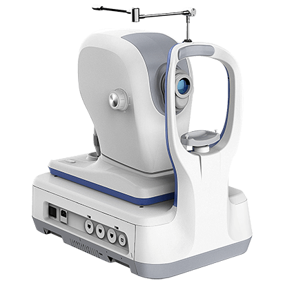
Optical Coherence Tomography (OCT) is an imaging method used to generate a picture of the back of the eye, called the retina. OCT uses light waves to take cross-section pictures of your retina. The OCT is an excellent way to visualize the different layers of the retina and optic nerve in the eye. OCT is routinely used during check-up of patients with glaucoma.

Color Fundus Retinal Photography uses a fundus camera to record color images of the condition of the interior surface of the eye, in order to document the presence of disorders and monitor their change over time.

A fundus camera or retinal camera is a specialized low power microscope with an attached camera designed to photograph the interior surface of the eye, including the retina, retinal vasculature, optic disc, macula, and posterior pole (i.e. the fundus).

Edit Content

Slit Lamp

The binocular slit-lamp examination provides a stereoscopic magnified view of the eye structures in detail, enabling anatomical diagnoses to be made for a variety of eye conditions. A second, hand-held lens is used to examine the retina. A slit-lamp exam is usually done during a regular checkup with your eye doctor before the cataract surgery procedure.

Edit Content

NC Tonometer

NC Tonometer is used to perform Tonometry. Tonometry is a quick and simple test that checks the pressure inside your eyes. The results can help your doctor see if you're at risk for glaucoma. The pressure inside your eye is called intraocular pressure (IOP).

Edit Content

Applanation Tonometer

This test measures fluid pressure in your eye. The test involves using a slit lamp equipped with forehead and chin supports and a tiny, flat-tipped cone that gently comes into contact with your cornea. The test measures the amount of force needed to temporarily flatten a part of your cornea.

Edit Content

20D/90D LENS

This lens provides ultra resolution with radinal image with the binocular indirect ophthalmoscope during clinical practice or in the operating room.

Edit Content

DIRECT / INDIRECT OPHTHALMOSCOPE

Ophthalmoscopy is a test that look at the back of the eye called the fundus. The fundus consists of the retina, optic disc and blood vessels.

A direct ophthalmoscope is a device that produces an unreversed or upright image of around 15 x magnification.

An indirect ophthalmoscope produces a reversed or inverted image with 2 to 5 x magnification.

Edit Content

OPTICAL COHERENCE TONOGRAPHY (OCT)

Optical Coherence Tomography (OCT) is an imaging method used to generate a picture of the back of the eye, called the retina. OCT uses light waves to take cross-section pictures of your retina. The OCT is an excellent way to visualize the different layers of the retina and optic nerve in the eye. OCT is routinely used during check-up of patients with glaucoma.

Edit Content

FUNDUS CAMERA - CANON

Color Fundus Retinal Photography uses a fundus camera to record color images of the condition of the interior surface of the eye, in order to document the presence of disorders and monitor their change over time.

A fundus camera or retinal camera is a specialized low power microscope with an attached camera designed to photograph the interior surface of the eye, including the retina, retinal vasculature, optic disc, macula, and posterior pole (i.e. the fundus).

FAQ

Frequently asked questions

Inflammation or swelling in any of the part of the uveal tract is called uveitis. Depending on the location of inflammation, it is called anterior uveitis (iris and ciliary body is involved), intermediate uveitis (ciliary body and the vitreous is involved), posterior uveitis (choroids and retina involved), panuveitisd (all layers of the uveal tract are involved).

Causes of Uveitis are many.

  • It may result from an infection (bacteria like tuberculosis, viruses like herpes, parasites like toxoplasmosis)
  • It may also be related to an autoimmune disease (with or without involvement of other parts of the body). This essentially is when our immune system recognizes a part of our own body as foreign and elicits a reaction in the form of uveitis. The uveitis may be the presenting manifestation of an underlying systemic disease.
  • It can occur as a result of injury to the eye.
  • It can occur due to causes within the eye like cataract and retinal detachment
  • Sometimes the cause is not known at the first instance. As we follow-up the patient over a period of time, we may be able to pick up new clues or signs suggestive of a systemic disease. In such cases, they are labeled as idiopathic (cause not known) at the initial visit.

.

Depending on the structure involved, the symptoms can vary from

  • Pain
  • Redness
  • Inability to see light (photophobia)
  • Black spots moving in front of the eye
  • Blurring or reduction in vision

Nehrunagar Branch

1, Tapovan Society Nr. Nehrunagar Cross Road Satellite, Ahmedabad, Gujarat - 380015 | TEL: +91 7926753534

Relief Road Branch

Opp. Z. K. Hotel Relief Road, Ahmedabad, Gujarat - 380001 | TEL: +91 7925506257

Isanpur Branch

307-311, Iconic Isanpur, Near Honda showroom, Next to Indian Oil Petrol Pump, Naroda Narol Highway, Isanpur, Ahmedabad, Gujarat - 382443 | TEL: +91 9979393783

Bopal-Ambli Branch

102,103, B Square 2, Vikram Nagar, Iscon-Ambli-Bopal Road, Ahmedabad, Gujarat - 380054 | TEL: +91 8160755086