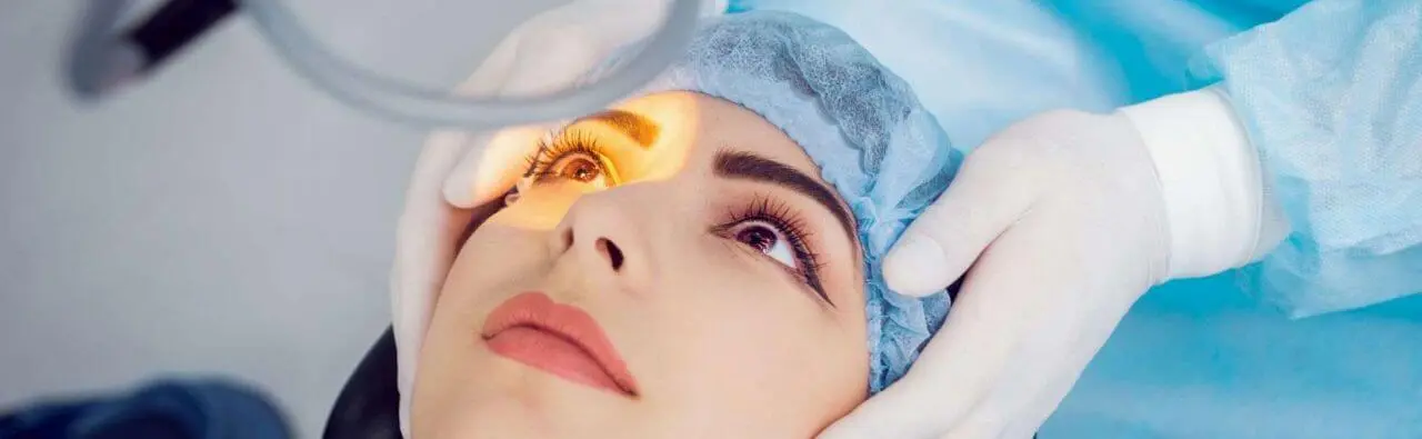
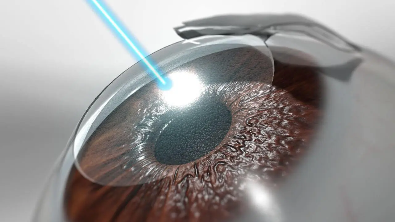

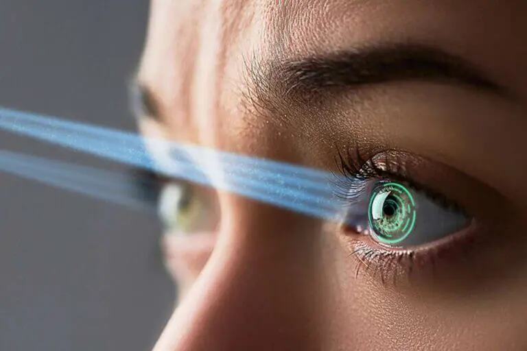
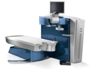
The World's Fastest Excimer Laser which minimizes dehydration of the corneal bed to increase treatment outcome accuracy. That means better vision. The fastest Eye Tracker available, moving 10 times faster than the eye is capable of to ensure the laser correction is always placed where it should be.
The binocular slit-lamp examination provides a stereoscopic magnified view of the eye structures in detail, enabling anatomical diagnoses to be made for a variety of eye conditions. A second, hand-held lens is used to examine the retina. A slit-lamp exam is usually done during a regular checkup with your eye doctor before the cataract surgery procedure.

NC Tonometer is used to perform Tonometry. Tonometry is a quick and simple test that checks the pressure inside your eyes. The results can help your doctor see if you're at risk for glaucoma. The pressure inside your eye is called intraocular pressure (IOP).

This lens provides ultra resolution with radinal image with the binocular indirect ophthalmoscope during clinical practice or in the operating room.

Ophthalmoscopy is a test that look at the back of the eye called the fundus. The fundus consists of the retina, optic disc and blood vessels.
A direct ophthalmoscope is a device that produces an unreversed or upright image of around 15 x magnification.
An indirect ophthalmoscope produces a reversed or inverted image with 2 to 5 x magnification.


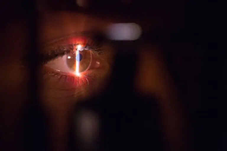
Corneal topography is a procedure used to monitor and measure changes that may occur to the shape and integrity of the cornea of your eye.

Optical Coherence Tomography (OCT) is an imaging method used to generate a picture of the back of the eye, called the retina. OCT uses light waves to take cross-section pictures of your retina. The OCT is an excellent way to visualize the different layers of the retina and optic nerve in the eye. OCT is routinely used during check-up of patients with glaucoma.
![]()
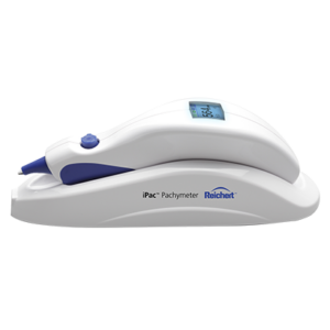
A pachymeter is a medical device used by the doctor to do a test called pachymetry. It is a simple, quick, painless test to measure the thickness of your cornea. With this measurement, your doctor can better understand your IOP reading, and develop a treatment plan that is right for your condition. The procedure takes only about few minutes to measure both eyes.