


The binocular slit-lamp examination provides a stereoscopic magnified view of the eye structures in detail, enabling anatomical diagnoses to be made for a variety of eye conditions. A second, hand-held lens is used to examine the retina. A slit-lamp exam is usually done during a regular checkup with your eye doctor before the cataract surgery procedure.

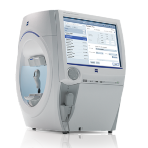
NC Tonometer is used to perform Tonometry. Tonometry is a quick and simple test that checks the pressure inside your eyes. The results can help your doctor see if you're at risk for glaucoma. The pressure inside your eye is called intraocular pressure (IOP).

This lens provides ultra resolution with radinal image with the binocular indirect ophthalmoscope during clinical practice or in the operating room.

Ophthalmoscopy is a test that look at the back of the eye called the fundus. The fundus consists of the retina, optic disc and blood vessels.
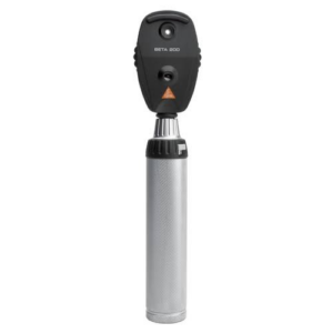
A direct ophthalmoscope is a device that produces an unreversed or upright image of around 15 x magnification.
An indirect ophthalmoscope produces a reversed or inverted image with 2 to 5 x magnification.

A visual field test measures how far the eye sees in any direction without moving and how sensitive the vision is in different parts of the visual field. This helps doctors to find certain types of injuries and disease, like glaucoma
Optical Coherence Tomography (OCT) is an imaging method used to generate a picture of the back of the eye, called the retina. OCT uses light waves to take cross-section pictures of your retina. The OCT is an excellent way to visualize the different layers of the retina and optic nerve in the eye. OCT is routinely used during check-up of patients with glaucoma.
![]()
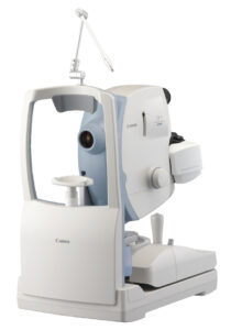
Color Fundus Retinal Photography uses a fundus camera to record color images of the condition of the interior surface of the eye, in order to document the presence of disorders and monitor their change over time.
A fundus camera or retinal camera is a specialized low power microscope with an attached camera designed to photograph the interior surface of the eye, including the retina, retinal vasculature, optic disc, macula, and posterior pole (i.e. the fundus).
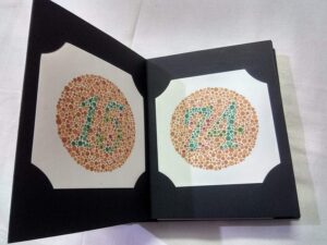
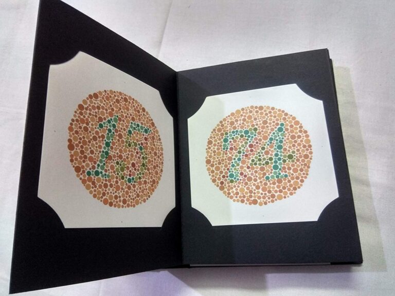
It measures your ability to tell the difference among colors. If you don't pass this test, you may have poor color vision, or your doctor may tell you that you're color blind.